
Prof. Shahzad Shams presently works as Head and Professor of the Neurosurgery Department at King Edward Medical College Lahore, Pakistan.
Pituitary Tumors / Sellar & Parasellar Tumors:
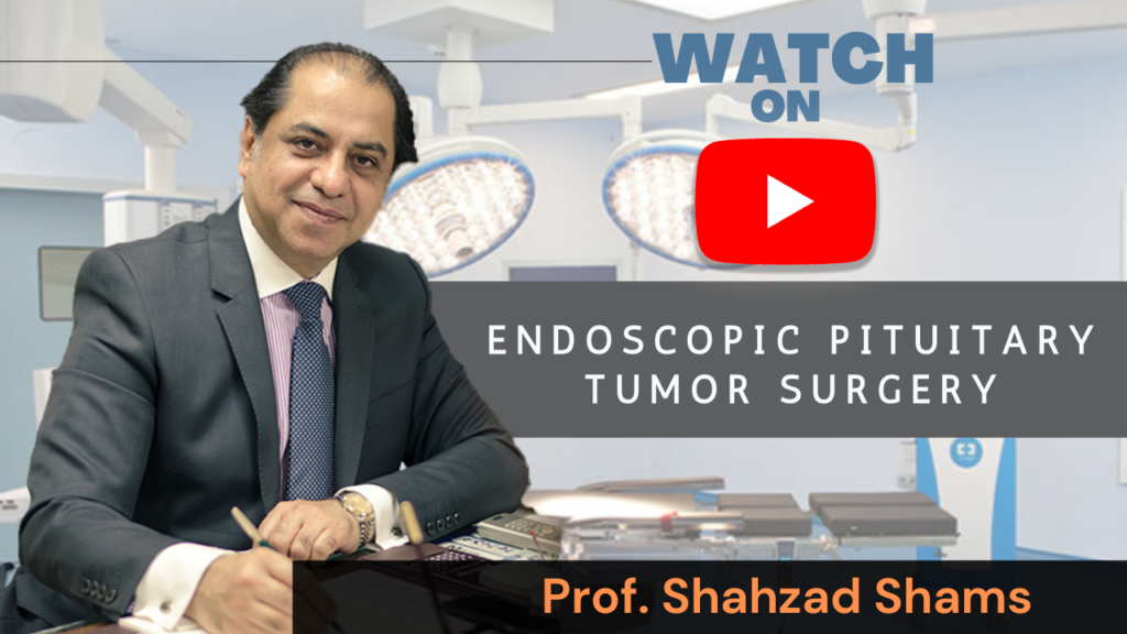
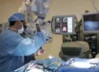
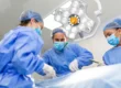
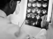
Professor Shahzad Shams is known the world over for providing Quality care to Pakistani and patients from abroad for Nasal technique of removal of brain tumors like Pituitary adenoma, Prolactinoma, Craniopharyngioma, Rathke’s cleft cyst, Sellar and Parasellar tumors using Transnasal Transsphenoidal endoscopic pituitary surgery technique of removal of the tumor through the nose also known as Transnasal endoscopic surgery of Pituitary tumors also called Transsphenoidal surgery.
Patients come from countries like US United States of America USA, United Kingdom UK Britain England, Canada, Italy, Greece, Germany, Spain, Australia, Saudi Arabia, United Arab Emirates UAE, Dubai, Kuwait, Qatar, Bahrain and Afghanistan requiring Neuro, Neurological Surgery or Neurosurgery management.