
Prof. Shahzad Shams presently works as Head and Professor of Neurosurgery Department at Lahore General Hospital, LGH, Lahore.
Intracranial Tuberculomas
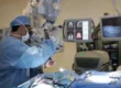
Best treatment of Brain Tuberculoma is Stereotactic biopsy which is Minimally invasive keyhole surgery which is safest surgery and done while patient is awake and stay in Hospital is only for 24 hours. On positive biopsy report of tuberculoma treatment with Anti tuberculous therapy completely cures the patient.
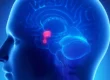
An intracranial tuberculoma originates by hematogenous spread from tuberculous lesions in other parts of the body, especially the lung. Tuberculomas are frequently multiple and are predominantly located in the posterior fossa in children and young adults, but may occur throughout the cerebral hemispheres.
The clinical presentation is similar to an intracranial tumor, with features of raised intracranial pressure, focal neurological signs and epileptic seizures, systemic symptoms of tuberculosis, such as fever, excessive perspiration and lethargy, occur in less than 50% of cases.
The MRI and CT scan appearance of a tuberculous granuloma is an area of low attenuation with a contrast-enhancing capsule. There is usually surrounding oedema and the lesions may be multiple. The tuberculoma is occasionally calcified.
Latest Advanced Treatment of Intracranial Tuberculomas
Best treatment of Brain Tuberculoma is Stereotactic biopsy which is minimally invasive keyhole safest surgery and done while patient is awake and stay in Hospital is only for 24 hours. On positive biopsy report of tuberculoma treatment with Anti tuberculous therapy completely cures the patient.